A radiology report follows a standard format: patient and exam details, clinical history, technique, comparison, findings, impression and the radiologist's sign-off. The impression is the part doctors read first and patients find hardest. Below is each section, a filled chest X-ray sample, and the same report rewritten in plain language.
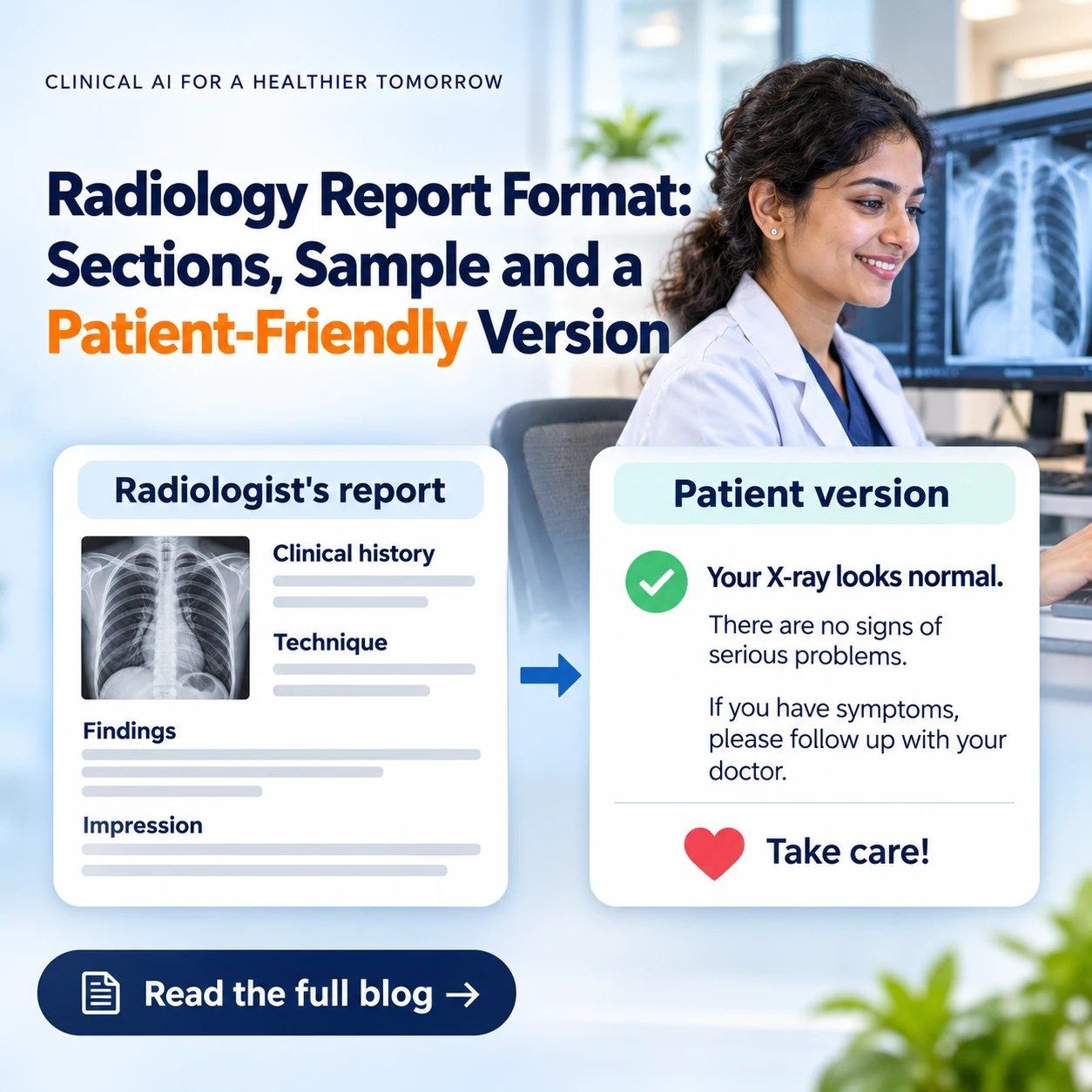
The Standard Radiology Report Format at a Glance
| Section | What goes in it | Example |
| Patient and exam details | Name, age, sex, patient ID, referring doctor, exam name, date and time, centre | 45/M, X-ray chest PA view, 18-11-2026 |
| Clinical history | Why the scan was ordered | Cough for 2 weeks |
| Technique | Views, sequences and any contrast used | Single PA view in full inspiration |
| Comparison | Earlier scans compared, or "none available" | None available |
| Findings | What the radiologist sees, structure by structure | Both costophrenic angles are clear |
| Limitations | Anything that reduces accuracy, when relevant | Study limited by patient movement |
| Impression | The conclusion: a diagnosis or a short list of possibilities | No acute abnormality |
| Recommendations | Follow-up or further tests, when appropriate | Repeat X-ray after treatment |
| Sign-off | Radiologist's name, qualification, registration number, signature, date and time | Dr [name], MD (Radiology) |
This order follows the American College of Radiology's practice parameter on communicating imaging findings. RSNA's free RadReport library has ready templates for many common scans.
How to Write Each Section Well
- Clinical history: Write the question the referring doctor is asking, not just "for evaluation". The ACR guideline says the report should answer any specific clinical question, and say so when it cannot.
- Technique: Name the views or sequences, and any contrast or medication given. If the patient had a reaction, record it.
- Comparison: Always say whether earlier scans were compared. "None available" is better than a blank, because the next radiologist needs to know.
- Findings: Go structure by structure in a fixed order, so nothing is skipped and readers know where to look. Include normal findings that answer the clinical question, such as "no pleural effusion" in a patient with breathlessness.
- Impression: Lead with the most important finding. Give a specific diagnosis when possible and a short list of possibilities when not. Suggest a follow-up test when it would settle the question.
- Sign-off: The reporting radiologist's name, qualification and registration number, with the date and time of the final report.
Common Mistakes in Radiology Reports
- An impression that repeats every finding instead of concluding
- "Cannot be ruled out" with no suggested next step
- No comparison line
- Abbreviations such as CP angles, BVM or HSM that patients then search online
- A template left unedited, such as a normal gall bladder described in a patient who has had it removed
How the Format Changes by Scan Type
| Scan | The technique section usually notes | Findings usually follow this order |
| X-ray | Views (PA, AP, lateral) and patient position | Lungs, pleura, heart, mediastinum, diaphragm, bones, soft tissues |
| Ultrasound (USG) | Approach and the organs scanned | Organ by organ (liver, gall bladder, bile ducts, pancreas, spleen, kidneys, bladder), with sizes |
| CT | Plain or contrast, and which phases | Region by region, with measurements of anything abnormal |
| MRI | Sequences (such as T1, T2, FLAIR, diffusion) and contrast | Structure by structure, with the signal on each sequence |
Sample: Chest X-ray Report
This sample is for illustration only. The patient is fictional.
| Field | Report |
| Patient | Ramesh Kumar, 45/M, ID 000123 |
| Referred by | Dr A. Mehta |
| Exam | X-ray chest, PA view, 18-11-2026, 10:40 am |
| Clinical history | Cough for 2 weeks. No fever. |
| Technique | Single PA view in full inspiration. |
| Comparison | None available. |
Findings
- Prominent bronchovascular markings in both lower zones.
- No consolidation or collapse.
- Both costophrenic angles are clear.
- Cardiac size is within normal limits.
- Mediastinum and both hila are normal.
- Both domes of the diaphragm are normal.
- Bony thorax and soft tissues are normal.
Impression: Prominent bronchovascular markings in both lower zones, which may represent bronchitis. Clinical correlation suggested.
Reported by: Dr [name], MD (Radiology), Reg. No. [number], 18-11-2026, 12:15 pm
The Same Report, Rewritten for the Patient
The patient-friendly version sits alongside the radiologist's report and does not change it. Each term gets one plain sentence, and the patient gets a next step.
| The report says | What it means for you |
| PA view | The X-ray was taken from back to front, the standard way for a chest X-ray. |
| Prominent bronchovascular markings in both lower zones | The airways and blood vessels in the lower part of both lungs show up more than usual. This is common and not specific to one disease. It can be seen with bronchitis, a recent chest infection or smoking. |
| No consolidation or collapse | There is no sign of pneumonia, and no part of the lung has collapsed. |
| Costophrenic angles are clear | There is no sign of fluid around the lungs. |
| Cardiac size is within normal limits | Your heart is a normal size. |
| May represent bronchitis | The X-ray may fit with bronchitis, which means irritated, swollen airways. |
| Clinical correlation suggested | Your doctor will match this X-ray with your symptoms and examination before deciding on treatment. It does not mean the X-ray is unclear. |
What to do next: Show this report to the doctor who asked for the X-ray. See a doctor sooner if you become breathless or have chest pain or a high fever.
Nothing was removed or softened. The patient simply no longer has to search "bronchovascular markings" at midnight. This sample is general information and is not medical advice. If you are a patient trying to read other test results, see our guide on how to read a medical lab report.
Why Imaging Centres Are Adding a Patient-Friendly Version
Patients often read their reports before they see their doctor, because many centres send them on WhatsApp, email or a portal. A report written only for doctors leaves them searching terms online or calling the centre. The evidence so far is encouraging, with some limits:
- Less anxiety: In a 2023 JACR survey of 1,033 patients at 10 outpatient imaging centres in Denver, 70% said the patient-friendly report reduced their anxiety about the results, 81% felt better prepared, and 87% said conversations with their doctor were more productive (Radiology Business). Respondents had viewed the report and agreed to be contacted, so the results may lean towards patients who liked it.
- Better understanding: In a 2026 Pediatric Radiology study at Stanford and Duke children's hospitals, 92.6% of families who answered said the patient-friendly report improved their understanding of the results (AuntMinnie). Understanding was self-reported rather than tested.
- Delivery matters: In the same study, only 9% of families opened the patient-friendly report when it was a separate item in the portal, against 50.5% for the standard report. The plain-language version works best inside the report patients already open.
The JACR authors also noted that clearer reports could ease the load of imaging-related phone calls to the centre.
Want patients to understand their scan before they see their doctor? NirogGyan works with diagnostic centres to turn static reports into patient-friendly smart reports.
Explore for labs and centresNew to imaging software? Read What is PACS?, how RIS improves workflow and how modern PACS can improve patient understanding.
Frequently Asked Questions
What are the main sections of a radiology report?
Patient and exam details, clinical history, technique, comparison, findings, impression and the radiologist's sign-off. Many reports also note limitations and recommendations for follow-up.
What is the difference between findings and impression in a radiology report?
Findings describe what the radiologist sees, structure by structure. The impression is the conclusion: the most likely diagnosis or a short list of possibilities, plus any suggested next step. Doctors usually read the impression first.
What does "clinical correlation" mean in a radiology report?
It asks the referring doctor to interpret the scan together with the patient's symptoms, examination and other test results. It does not mean the scan is faulty, and on its own it does not mean something serious was found.
Is there a standard radiology report format in India?
Indian centres commonly follow the internationally used structure: details, history, technique, comparison, findings and impression, as set out in the American College of Radiology's reporting guideline. Free templates for many scans are available in RSNA's RadReport library.
Can a radiology report be rewritten for patients?
Yes. A patient-friendly version explains each finding in plain language and says what happens next, while the radiologist's original report stays unchanged. In a 2023 survey of 1,033 patients at US imaging centres, 70% said such a report reduced their anxiety.
Sources
- ACR Practice Parameter for Communication of Diagnostic Imaging Findings (revised 2020)
- RSNA RadReport reporting templates
- Consumer-oriented radiology reports a "promising" strategy, Radiology Business, on the 2023 JACR survey
- Patient-friendly radiology reports improve family comprehension, AuntMinnie, August 2026